Lowering Costs, Saving More Lives
Advances in reconstructive surgery benefit not only patients and their families, but the community as a whole
Congenital defects like a cleft palate. After-cancer treatment related to tumor removal. Trauma as a result of a car accident. To repair these conditions and more, reconstructive surgery is vital and oftentimes lifesaving. And advances in reconstruction in recent years, such as 3D printing and skin substitutes, have led to transformative benefits for patients and the medical community.
“3D-imaging technology helps guide the operations, with the idea of making surgery shorter and more efficient,” says Rizal Lim, MD, plastic surgeon at the Burn and Reconstructive Centers of America in Augusta. That means patients need fewer additional surgeries, he says.
The benefits are remarkable. A cutting-edge therapy can not only make a person’s life better and reduce suffering and mortality, but it also saves money.
“A lot of medical procedures and treatments are very costly, so if we look at science as a way to improve our outcomes, as well as reduce our costs, that is a good value proposition to everyone,” says Steven Goudy, MD, director of Otolaryngology at Children’s Healthcare of Atlanta and Emory University.
Technology’s Growing Role
The technology behind 3D printing dates back to the mid-1980s, but its application in the medical field didn’t really begin until the early- to mid-1990s.
At that time, it was just used to make models for planning and surgical decision-making, says Scott J. Hollister, Patsy and Alan Dorris chair in Pediatric Technology in the Wallace A. Coulter Department of Biomedical Engineering at the Georgia Institute of Technology and Emory University. Now it’s also used to create devices that go into patients’ bodies, and even to print cells in gel materials that could eventually create organs.
“Printing of implantable devices started around 2010, and the majority of applications in this area right now are with metal, printing titanium for orthopedic and craniofacial reconstruction purposes,” says Hollister, who also directs the Center for 3D Medical Fabrication and the Tissue Engineering and Mechanics Laboratory at Georgia Tech.
Dr. Lim, who has worked at the Burn and Reconstructive Centers of America for about six years, uses 3D imaging technology in surgical planning for head and neck surgery at the acute care hospital.
“We can do cutting guides for the surgical planning and [make] surgical splints and intra- oral splints for the teeth,” Dr. Lim says. “We can also have them custom-print 3D plates, which makes everything more efficient when we are doing these multilevel facial cases.”
Using 3D printing for surgical planning also helps decrease the overall amount of time someone is in surgery.
And while the majority of medical 3D printing is used to make models, Hollister’s team does print implantable devices. To date, the devices have been used in more than 30 patients.
These devices are all designed specifically for the individual. “We can readily take a patient’s data image and build a device from that,” Hollister says. “And the nice thing about 3D printing is [that] you cannot only make complex structures, but it’s very cost-effective to make patient-specific devices that you can’t do with other manufactured technologies.”
In 2018, Hollister’s team of biomedical engineers helped create three custom-made splints for Children’s Healthcare of Atlanta to assist the breathing of a seven-month-old battling a life-threatening airway obstruction – the first procedure of its kind in Georgia.
The engineers have also worked closely with ENT surgeons at Children’s Healthcare of Atlanta to print for craniofacial reconstruction, specifically noses, ears and a temporomandibular joint, which is the hinge joint between the temporal bone and the lower jaw.
“Your face is what’s visible to the world,” Dr. Goudy says. “So, if you have a different [facial structure] and/or are born with part of your face appearing different – you have a tumor or cancer, or there’s something that needs to be replaced or wasn’t there at birth – it has a visible, functional and psychological consequence.”
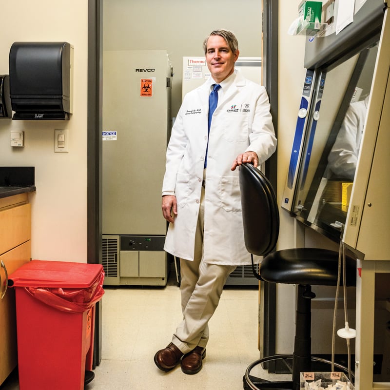
Typically, Dr. Goudy and his colleagues have “borrowed” different parts of a patient’s body to reconstruct an area, such as a recent 12-hour surgery in which part of a patient’s fibula (one of the bones in the lower leg) was removed and used to make an upper jaw.
Goudy says he’d like to be able to take “something that is off the shelf” that wouldn’t cause the complications or side effects of “borrowing” another body part. “Ideally, that implant or therapy would also tell or encourage the body to rebuild itself in the way that meets or addresses the needs of the patient,” he says.
Dr. Goudy’s lab at Emory University is also focused on regenerative therapies for children – to reconstruct or regrow bone, as well as tissue, using different models.
“There are not many regenerative or reconstructive options available, so I have a lab where we have models where we are actually using repurposed, FDA-approved drugs [currently used for multiple sclerosis] to improve wound healing in the mouth,” Dr. Goudy says. “The way that it functions is that we deliver it to a local area and it reduces the inflammatory part of wound healing and encourages tissue regeneration, and we’ve seen great success with that.”
His team is using pediatric bone cells in the lab to develop a system to deliver a specific protein called Jagged 1, using human cells to ultimately induce (or sort of teach) the body’s cells to regenerate bone.
Harsha Ramaraju and Ryan Akman, both researchers on Hollister’s team, are working on a project with the use of something called shape-memory material, and they have invented a material called poly(glycerol dodecanedioate).
“They developed a scheme to 3D print it using a variety of technologies,” he says. “Shape memory means you can print it in one shape, warm it up and sort of fold it into a different shape. We can deliver it into the body, and the body temperature can help it retake its original shape.”
While the material is still being studied, it’s shown promising results in pig arteries. “It’s good for cardiovascular and soft tissue applications,” Hollister adds. “It’s also resorbable, so it’ll go away in six months, and you can attach lots of different biological factors to it.”
Improving Cleft Lip Surgery
Heather Koehn, MD, pediatric ENT, facial plastic surgeon at Children’s Hospital of Georgia in Augusta and assistant professor in the Department of Otolaryngology at the Medical College of Georgia at Augusta University, primarily works with patients with craniofacial defects like cleft lip and cleft palate or children born with a small jaw.
Cleft lip is one of the most common birth defects in the U.S. Advancements in cleft lip surgery include a pre-surgical orthopedic therapy called nasoalveolar molding, or NAM, which uses custom-made devices to help narrow the cleft.
“It helps bring the edges of the cleft together. That’s from the bone of where the teeth come out in the gums,” Dr. Koehn says. “It also helps to bring the lip edges closer together.”
But what it does more than anything else – and what Dr. Koehn thinks makes the difference for so many kids – is to reshape the nose.
“We talk about cleft lip as a defect in the lip or a defect in the gums, but the nose is so severely affected in most of these kids, even when they have a minor form of a cleft lip,” she says. “NAM has helped cut down on how extensive the initial surgery has to be and also potentially avoids the need for future nasal surgeries.”
NAM isn’t always an option in advance of cleft lip surgery: A medical team needs to be able to collaborate with a dentist or orthodontist, which is not always possible. However, cleft-lip surgeons at the Children’s Hospital of Georgia have the unique opportunity to partner with the Dental College of Georgia at Augusta University to bring this state-of-the-art technology to their patients.
Hope for Burn and Wound Survival
When David Herndon, MD, and CEO of the Joseph M. Still Research Foundation in Augusta, began working as a burn doctor in the 1970s, half the young people who received burns covering 50% of their bodies died. Thanks to advances in medicine and research over the past 40 years, now even a 98% total body burn may be survivable.
“Probably the greatest single advance in this period was a switch from using topical creams and goos on burns over long periods of time to an early surgical removal of the burn wound and coverage with [grafted] skin from other people,” Dr. Herndon says. At first, that meant using skin from cadavers. Now, Dr. Herndon says there have been other major advances in developing skin substitutes that could be used instead of cadaver skin, which gives better cosmetic results.
Dr. Herndon, who has collaborated with the Burn and Reconstructive Centers of America for 30 years, literally wrote the textbook on burn care – Total Burn Care – which is the leading manual on burns around the world.
Over the past four decades, he’s worked with a lot of skin substitutions in burn and wound care, most notably one that is part shark skin with a matrix of collagen from cows.
“This two-layer product is applied to surgically prepared wounds,” says Dr. Herndon. “The bottom layer is made from collagen from cows, with a component from sharks that helps maintain openings in the dressing that encourage ingrowth of the patient’s own cells that populate the dressing. The outer layer is a silicone that keeps water in and bacteria out and allows oxygen to penetrate to the wound. The plastic is removed after three weeks and the inner collagen layer is skin-grafted. The results are cosmetically superior.”
Doctors are also now able to grow a patient’s own skin from a small piece of tissue, a process called tissue-culture growth of skin. A piece of skin the size of a silver dollar can be grown in a laboratory for skin grafts, and within three to five weeks, it can cover the whole body, he says.
Another development is a product called ReCell. It is a spray-on skin system, using a patient’s own skin cells, to treat skin loss, scarring and depigmentation after a burn injury.
“It’s been successful in improving rates of wound healing in deep, second-degree burns and very large burns,” says Dr. Herndon.
And there is much more on the horizon for helping improve burn and wound care, as well as reconstructive surgery techniques to reduce scars by bringing new tissues into the affected area. “Advances in all of these areas have led to the tremendous diminishment of mortality and improvement of morbidity over time,” says Dr. Herndon.
Rajiv Sood, MD, chief of Plastic and Reconstructive Services at the Burn and Reconstructive Centers of America, and Dr. Herndon’s colleague, says the center is also focused on scar resurfacing, laser resurfacing and skin substitution.
“We still do tissue expansion with normal skin to advance it and remove some of the burned skin, but one of the biggest advances is that we are thinking about this more and more,” he adds. “As our survival rates across the country have improved, we are now thinking about how we improve function. And, more importantly, how these patients reintegrate into society.”